Jessica Auyeung, Monica Lee
Stevens–Johnson syndrome (SJS) and toxic epidermal necrolysis (TEN) are rare, severe idiosyncratic reactions characterized by extensive necrosis and detachment of the epidermis, commonly triggered by medications. The annual incidence is 1 or 2 cases per million.1 There is usually a prodrome of malaise and fever, followed by rapid onset of erythematous or purpuric macules and plaques that progress to epidermal necrosis and sloughing. SJS and TEN are considered to lie along the same spectrum of disease, differing only in terms of how much of the skin surface is affected. If less than 10% of the body surface is affected, the diagnosis is SJS, and if more than 30% is affected, the diagnosis is TEN; if between 10% and 30% is affected, the diagnosis is SJS–TEN overlap. Although medications such as allopurinol, antibiotics, and antiepileptics are the leading causes in most cases, infections, vaccines, and food have been implicated.1,2 Although fluoroquinolone- associated SJS/TEN has been reported in the literature, the number of cases due to this group of drugs has been relatively small compared with cases due to other drugs. Systemic corticosteroids and immunosuppressive therapies have been used in the management of SJS/TEN, but high-quality evidence supporting their use is lacking.
We describe a probable case of ciprofloxacin-induced SJS treated with methylprednisolone, prednisone, and cyclosporine.
A 66-year-old woman presented to the emergency department of a tertiary care hospital with a 2-day history of rash that had developed on her trunk and spread to her neck, arms, and legs.* Her other symptoms were fever, progressive dysphagia, odynophagia, dysuria, and painful ulcerations to her oral and genital mucosa. The Nikolsky sign was positive (i.e., application of mechanical pressure to the skin resulted in epidermal detachment), which indicated necrolysis.1 The patient was febrile, with a temperature of 38.2°C. Her blood pressure was 116/66 mm Hg, heart rate 93 beats/min, respiratory rate 20 breaths/min, and oxygen saturation 100% on 2 L/min via nasal prongs. Her past medical history included chronic obstructive pulmonary disease, osteoporosis, osteoarthritis with left total hip replacement, stress urinary incontinence, and migraines. She had allergies to sulfonamides and azithromycin, with a reported reaction of rash to both agents. She also reported abdominal pain associated with acetaminophen compound with codeine. Eight days before the admission, she had undergone a urethral sling procedure, and was started on ciprofloxacin 500 mg orally twice daily for 10 days as prophylaxis for urinary tract infection. Her other long-term home medications were fluticasone– salmeterol, salbutamol, tiotropium, ipratropium, quetiapine, and risedronate.
The results of laboratory investigations on admission were unremarkable. The leukocyte count was 9.6 × 10⁹/L, and eosinophils were not detected. C-reactive protein and erythrocyte sedimentation rate were not checked at the time of admission, but 2 days after admission, C-reactive protein was 110.1 mg/L and erythrocyte sedimentation rate was 85 mm/h. The patient was admitted with a working diagnosis of SJS. Skin biopsy showed transepidermal necrosis with perivascular chronic inflammation, and the morphologic findings were consistent with SJS/TEN.
Methylprednisolone 50 mg IV every 12 h was initiated. Supportive therapy included morphine, lubricating eye drops, and mucositis mouthwash (consisting of nystatin and lidocaine). Her preadmission medications were continued, with the exception of ciprofloxacin. On day 3 of the admission, a dermatologist initiated cyclosporine 3 mg/kg IV daily as adjunctive treatment. The patient weighed 66 kg and therefore received cyclosporine 195 mg daily. At 3.5 weeks, the cyclosporine therapy was converted to oral administration, and at 1 month, it was tapered to 2 mg/kg for 4 days, then 1 mg/kg for 7 days. Cyclosporine was discontinued on day 44 of the hospital stay. After 12 days of IV methylprednisolone, the corticosteroid therapy was stepped down to oral prednisone 50 mg daily. After 1 week, the prednisone was tapered to 40 mg for 1 day and then 20 mg the following day; the dose was then reduced by 5 mg every 5 days. At the time of discharge (2 months after admission), 5 days of prednisone therapy remained to be completed.
Improvement to her skin rash became apparent 1 week after initiation of cyclosporine. Skin sloughing and formation of bullae halted after 1 month of therapy. However, other complications prolonged the patient’s hospital stay. The most significant complication was continued dysphagia and odynophagia because of severe esophagitis and ulceration, as seen on endoscopy. These problems were thought to be related to the SJS. The patient was unable to tolerate oral intake and required total parenteral nutrition for 40 days. Other complications included significant pain, a urinary tract infection, and thrombosis related to the central catheter.
With respect to therapeutic drug monitoring, serum cyclosporine trough concentrations were monitored periodically during the course of IV therapy to ensure the drug did not reach toxic levels. The trough concentration never exceeded 286 μg/L.
The patient was discharged home after a 2-month hospital stay, with full recovery of her skin and some residual dysphagia.
In this patient, it was presumed that ciprofloxacin, administered prophylactically following the urethral sling procedure, had caused the SJS. The Naranjo probability scale3 was used to assess the likelihood of the adverse reaction being due to this drug. The Naranjo score was 6/13, indicating a probable adverse drug reaction associated with ciprofloxacin. Points were assigned for previous conclusive reports of this adverse reaction (+1), appearance of the adverse event after administration of the medication (+2), no alternative cause that on its own could have caused the reaction (+2), and confirmation by objective evidence (+1). Rechallenge with ciprofloxacin, administration of a placebo, and measurement of serum ciprofloxacin concentrations were not performed. It was also unknown whether the patient had ever received ciprofloxacin previous to this encounter, or whether the severity of her adverse reaction would have changed with adjustment of the ciprofloxacin dose. Also, the SJS did not improve when the drug was discontinued (although it improved subsequently, after initiation of treatment). Eleven other cases involving ciprofloxacin-induced SJS or TEN were identified in the literature.4–13 Most of the cases were treated with supportive care only, with systemic corticosteroids being used in 5 cases.4,6,9,10,12 In one case, the patient was treated with systemic corticosteroids and tacrolimus.12
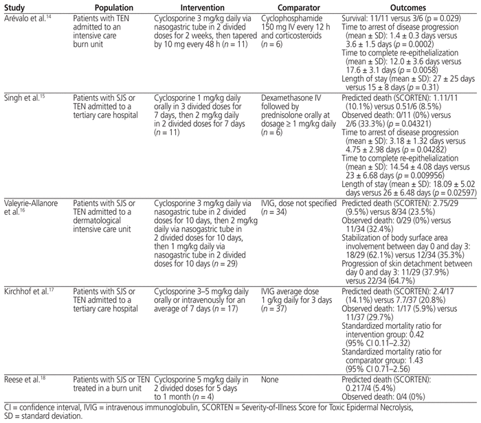
Although the use of systemic corticosteroids in SJS or TEN is common in clinical practice, there is a lack of strong evidence for their use.1 The rationale for using immunosuppressive agents is that they may suppress the cytotoxic reaction that results in keratinocyte apoptosis, the theorized pathophysiologic process of SJS/TEN. Given the lack of evidence for benefit of corticosteroids, consideration of alternative therapies, such as cyclosporine, is warranted. The studies evaluating cyclosporine for the management of SJS/TEN are summarized in Table 1.14–18
Table 1 Summary of Evidence for Using Cyclosporine to Treat Stevens–Johnson Syndrome (SJS) and/or Toxic Epidermal Necrolysis (TEN)
It is difficult to draw conclusions from these studies, which were a case series,18 a chart review,17 and open-label, uncontrolled studies using a variety of cyclosporine regimens and treatment durations.14–16 In most of these studies, individuals treated with cyclosporine monotherapy were compared with historical controls treated with other therapies. No randomized controlled trials were identified in the literature, and given the rarity of TEN/SJS, recruiting a sufficient number of participants for such a trial would be challenging.
Three studies15–17 compared the observed mortality rate with the predicted mortality rate, as determined by the severity-of-illness score for toxic epidermal necrolysis (SCORTEN), a validated TEN-specific prognostic score,19 in the intervention and comparator groups. An observed mortality rate that is lower than the predicted rate implies treatment benefits. In all 3 studies, the observed death rate was lower than the predicted death rate in the cyclosporine group, but higher than predicted in the comparator group. These results suggest a survival benefit associated with cyclosporine treatment, which needs to be further explored with controlled clinical trials. Statistical analysis between the intervention and comparator groups was performed only by Arévalo and others14 and Singh and others.15 Both of these studies found significant differences in favour of cyclosporine in terms of survival, time to arrest of disease progression, and timing of re-epithelization of skin. Singh and others15 also found that the duration of hospitalization was significantly lower in the cyclosporine group relative to those who received corticosteroids, but Arévalo and others14 found no significant difference in length of stay between the 2 treatment groups.
With respect to safety, Singh and others15 described development of corneal ulceration in one patient in the cyclosporine group; no adverse effects were reported in the group that received systemic steroids. The authors attributed the observed adverse effect to inadvertent continued use of the offending drug in eye drop form. Arévalo and others14 compared cyclosporine with cyclophosphamide and corticosteroids, and found no significant difference in terms of sepsis (8/11 versus 5/6, p = 0.99) and overall organ failure (mean number of organs affected 1.1 versus 2.3, p = 0.11). However, the cyclophosphamide group had significantly more cases of organ failure in 4 or more organs (2/11 versus 3/6, p = 0.029) and significantly more cases of leukopenia (0/11 versus 4/6, p = 0.006). Valeyrie-Allanore and others16 did not discuss the occurrence of adverse effects in their comparison group, but among the 29 patients receiving cyclosporine, 3 had to stop therapy because of acute hallucinations that were suspected to be related to reversible posterior leukoencephalopathy, transitory neutropenia, and severe infection, respectively. Among the 26 individuals who completed treatment, adverse effects were increased blood pressure (n = 3), renal impairment (n = 2), and sensitive neuropathy (n = 1).
Very little information is available about therapeutic drug monitoring of cyclosporine in this setting, and there are no target concentrations for cyclosporine for this indication. Valeyrie-Allanore and others16 reported the performance of therapeutic drug monitoring to avoid toxicity.16 In the case reported here, trough cyclosporine concentration never exceeded the upper limit of the target concentration range as defined for solid organ transplant, which is 100–400 μg/L.20 Future studies should explore the optimal dose and duration of cyclosporine, and the utility of therapeutic drug monitoring.
This case illustrates further experience with the combination of cyclosporine and corticosteroids in the treatment of SJS. This combination could be considered for patients with SJS that is unresponsive to corticosteroids alone.
1 Creamer D, Walsh SA, Dziewulski P, Exton LS, Lee HY, Dart JKG, et al. UK guidelines for the management of Stevens-Johnson syndrome/toxic epidermal necrolysis in adults 2016. Br J Dermatol. 2016;174(6):1194–227.

2 Harr T, French LE. Severe cutaneous adverse reactions: acute generalized exanthematous pustulosis, toxic epidermal necrolysis and Stevens-Johnson syndrome. Med Clin North Am. 2010;94(4):727–42.
3 Naranjo CA, Busto U, Sellers EM, Sandor P, Ruiz I, Roberts EA, et al. A method for estimating the probability of adverse drug reactions. Clin Pharmacol Ther. 1981;30(2):239–45.
4 Tham TC, Allen G, Hayes D, McGrady B, Riddell JG. Possible association between toxic epidermal necrolysis and ciprofloxacin. Lancet. 1991;338 (8765):522.
5 Sakellariou G, Koukoudis P, Karpouzas J, Alexopoulos E, Papadopoulou D, Chrisomalis F, et al. Plasma exchange (PE) treatment in drug-induced toxic epidermal necrolysis (TEN). Int J Artif Organs. 1991;14(10):634–8.
6 Moshfeghi M, Mandler HD. Ciprofloxacin-induced toxic epidermal necrolysis. Ann Pharmacother. 1993;27(12):1467–9.
7 Win A, Evers ML, Chmel H. Stevens- Johnson syndrome presumably induced by ciprofloxacin. Int J Dermatol. 1994;33(7):512–4.
8 Livasy CA, Kaplan AM. Ciprofloxacin-induced toxic epidermal necrolysis: a case report. Dermatology. 1997;195(2):173–5.
9 Hallgren J, Tengvall-Linder M, Persson M, Wahlgren CF. Stevens-Johnson syndrome associated with ciprofloxacin: a review of adverse cutaneous events reported in Sweden as associated with this drug. J Am Acad Dermatol. 2003;49(Suppl 5):S267–9.
10 Jongen-Lavrencic M, Schneeberger PM, van der Hoeven JG. Ciprofloxacin-induced toxic epidermal necrolysis in a patient with systemic lupus erythematosus. Infection. 2003;31(6):428–9.
11 Mandal B, Steward M, Singh S, Jones H. Ciprofloxacin-induced toxic epidermal necrolysis (TEN) in a nonagenarian: a case report. Age Ageing. 2004;33(4):405–6.
12 Okan G, Yaylaci S, Peker O, Kaymakoglu S, Saruc M. Vanishing bile duct and Stevens-Johnson syndrome associated with ciprofloxacin treated with tacrolimus. World J Gastroenterol. 2008;14(29):4697–700. 
13 Upadya GM, Ruxana K. Toxic epidermal necrolysis and agranulocytosis: rare adverse effects of ciprofloxacin. Indian J Med Sci. 2009;63(10):461–3.
14 Arévalo JM, Lorente JA, González-Herrada C, Jimenez-Reyes J. Treatment of toxic epidermal necrolysis with cyclosporin A. J Trauma. 2000;48(3): 473–8.
15 Singh GK, Chatterjee M, Verma R. Cyclosporine in Stevens Johnson syndrome and toxic epidermal necrolysis and retrospective comparison with systemic corticosteroid. Indian J Dermatol Venereol Leprol. 2013;79 (5):686–92.
16 Valeyrie-Allanore L, Wolkenstein P, Brochard L, Ortonne N, Maîıtre B, Revuz J, et al. Open trial of ciclosporin treatment for Stevens–Johnson syndrome and toxic epidermal necrolysis. Br J Dermatol. 2010;163(4):847–53.
17 Kirchhof MG, Miliszewski MA, Sikora S, Papp A, Dutz JP. Retrospective review of Stevens-Johnson syndrome/toxic epidermal necrolysis treatment comparing intravenous immunoglobulin with cyclosporine. J Am Acad Dermatol. 2014;71(5):941–7.
18 Reese D, Henning JS, Rockers K, Ladd D, Gilson R. Cyclosporine for SJS/TEN: a case series and review of the literature. Cutis. 2011;87(1):24–9.
19 Bastuji-Garin S, Fouchard N, Bertocchi M, Roujeau JC, Revuz J, Wolkenstein P. SCORTEN: a severity-of-illness score for toxic epidermal necrolysis. J Invest Dermatol. 2000;115(2):149–53.
20 Cyclosporine. In: Lexi-drugs online [database on the Internet]. Hudson (OH): Lexi-Comp, Inc; [cited 2013 Oct 4]. Available from: http://online.lexi.com. Subscription required to access content.
*The patient provided verbal consent for publication of this case report. ( Return to Text )
Competing interests: None declared. ( Return to Text )
Funding: None received. ( Return to Text )
Canadian Journal of Hospital Pharmacy, VOLUME 71, NUMBER 4, July-August 2018